

 (Human Specific) Antibody - Western Blot")
 Antibody - Western Blot")

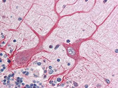
NOTCH 1 Antibody
Rabbit Polyclonal
$50.00 to US & $70.00 to Canada for most products. Final costs are calculated at checkout.
Background
Anti-Notch 1 Antibody recognizes Notch 1 that is synthesized in the endoplasmic reticulum as an inactive form which is proteolytically cleaved by a furin-like convertase (S1 cleavage) in the trans-golgi network before it reaches the plasma membrane to yield an active, ligand-accessible form. Cleavage results in a C-terminal fragment N(TM) and a N-terminal fragment N(EC). Following ligand binding, it is cleaved (S2 cleavage) by TNF-alpha converting enzyme (TACE) to yield a membrane-associated intermediate fragment called Notch extracellular truncation (NEXT). This fragment is then cleaved by presenilin-dependent gamma-secretase (S3 cleavage) to release the intracellular domain (NICD) from the membrane.
Product Details
Target Details
Application Details
Formulation
Shipping & Handling
This product is for research use only and is not intended for therapeutic or diagnostic applications. Please contact a technical service representative for more information. All products of animal origin manufactured by Rockland Immunochemicals are derived from starting materials of North American origin. Collection was performed in United States Department of Agriculture (USDA) inspected facilities and all materials have been inspected and certified to be free of disease and suitable for exportation. All properties listed are typical characteristics and are not specifications. All suggestions and data are offered in good faith but without guarantee as conditions and methods of use of our products are beyond our control. All claims must be made within 30 days following the date of delivery. The prospective user must determine the suitability of our materials before adopting them on a commercial scale. Suggested uses of our products are not recommendations to use our products in violation of any patent or as a license under any patent of Rockland Immunochemicals, Inc. If you require a commercial license to use this material and do not have one, then return this material, unopened to: Rockland Inc., P.O. BOX 5199, Limerick, Pennsylvania, USA.