Datasheet is currently unavailable. Try again or CONTACT US
Datasheet SDS
$665.00 /Per Item
Availability: Ships next business day
Shipping info:

$50.00 to US & $70.00 to Canada for most products. Final costs are calculated at checkout.

Background

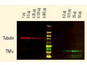
GFP Fluorescein Conjugated Antibody is designed for immunofluorescence microscopy, fluorescence based plate assays (FLISA) and fluorescent western blotting. This product is also suitable for multiplex analysis, including multicolor imaging, utilizing various commercial platforms.

Product Details

Anti-GFP (GOAT) Antibody Fluorescein Conjugated (Min X Hu Ms & Rt Serum Proteins) - 600-102-215
goat anti-GFP Antibody fluorescein Conjugation, FITC conjugated goat anti-GFP antibody, Green Fluorescent Protein, GFP antibody, Green Fluorescent Protein antibody, EGFP, enhanced Green Fluorescent Protein, Aequorea victoria, Jellyfish
Goat
Fluorescein (FITC)
Polyclonal
IgG

Target Details

GFP, eGFP, rGFP
Recombinant Protein
Recombinant Green Fluorescent Protein (GFP) fusion protein corresponding to the full length amino acid sequence (246aa) derived from the jellyfish Aequorea victoria.
GFP Fluorescein Conjugated Antibody was prepared from monospecific antiserum by immunoaffinity chromatography using Green Fluorescent Protein (Aequorea victoria) coupled to agarose beads followed by solid phase adsorption(s) to remove any unwanted reactivities.   Assay by immunoelectrophoresis resulted in a single precipitin arc against anti-Goat Serum, anti-Fluorescein and purified and partially purified Green Fluorescent Protein (Aequorea victoria).  No reaction was observed against Human, Mouse and Rat Serum Proteins.

Application Details

Dot Blot, WB
FC, IF, IHC, Multiplex  - View References
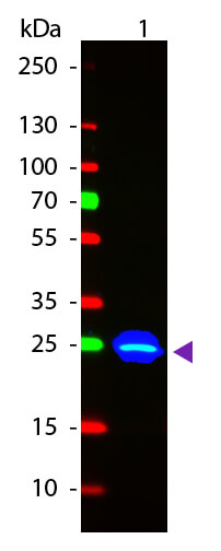
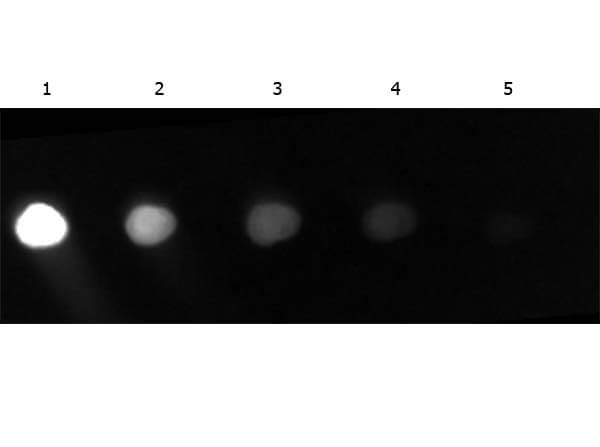
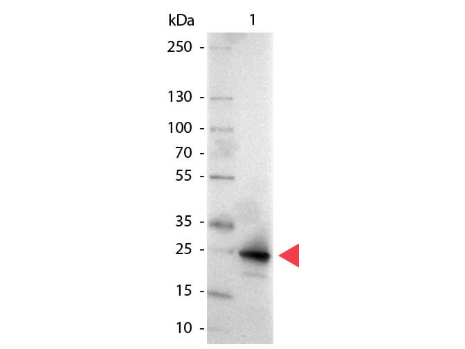
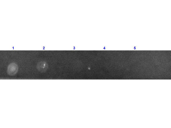
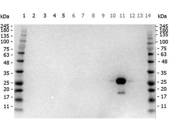
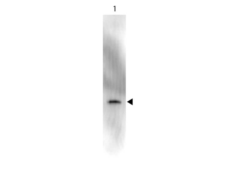
Anti-GFP Fluorescein Conjugated Antibody has been tested by dot blot and western blot and is suitable for immunomicroscopy and flow cytometry or FACS analysis as well as other antibody based fluorescent assays requiring lot-to-lot consistency.

Formulation

Lyophilized
1.0 mg/mL by UV absorbance at 280 nm
0.02 M Potassium Phosphate, 0.15 M Sodium Chloride, pH 7.2
0.01% (w/v) Sodium Azide
10 mg/mL Bovine Serum Albumin (BSA) - Immunoglobulin and Protease free
1.0 mL
Restore with deionized water (or equivalent)

Shipping & Handling

Ambient
Store anti-GFP at 4° C prior to restoration.   For extended storage aliquot contents and freeze at -20° C or below.  Avoid cycles of freezing and thawing.  Centrifuge product if not completely clear after standing at room temperature.  This product is stable for several weeks at 4° C as an undiluted liquid.  Dilute only prior to immediate use.
Expiration date is one (1) year from date of receipt.
(). Axon guidance at the spinal cord midline–a live imaging perspective. J Comp Neurol.
Applications
IF, Confocal Microscopy; IHC, ICC, Histology
(). TDP-43 proteinopathy alters the ribosome association of multiple mRNAs including the glypican Dally-like protein (Dlp)/GPC6. Acta Neuropathol Commun.
Applications
IF, Confocal Microscopy
(). Cables1 links Slit/Robo and Wnt/Frizzled signaling in commissural axon guidance. bioRxiv Preprint
Applications
IF, Confocal Microscopy
(). Identity and lineage fate of proteolipid protein 1 gene (Plp1)‐expressing cells in the embryonic murine spinal cord. Dev Dyn.
Applications
IHC, ICC, Histology
(). A novel distal convoluted tubule-specific Cre-recombinase driven by the NaCl cotransporter gene. Am J Physiol Renal Physiol.
Applications
IF, Confocal Microscopy
(). TF-ChIP Method for Tissue-Specific Gene Targets. Frontiers in Cellular Neuroscience
Applications
IHC, ICC, Histology; Multiplex Assay
(). Hypertonic Stress Causes Cytoplasmic Translocation of Neuronal, but Not Astrocytic, FUS due to Impaired Transportin Function. Cell Reports
Applications
IF, Confocal Microscopy
(). Brain Nat8l knockdown suppresses spongiform leukodystrophy in an aspartoacylase-deficient Canavan disease mouse model. Mol Ther.
Applications
IHC, ICC, Histology; Multiplex Assay
(). Controlling depth of cellular quiescence by an Rb-E2F network switch. Cell Rep.
Applications
FC, FACS, FLOW
(). 53BP1 and USP28 mediate p53-dependent cell cycle arrest in response to centrosome loss and prolonged mitosis. Elife
Applications
IF, Confocal Microscopy
(). Tumor necrosis factor receptor superfamily costimulation couples T cell receptor signal strength to thymic regulatory T cell differentiation. Nat Immunol.
Applications
FC, FACS, FLOW
(). CEP162 is an axoneme-recognition protein promoting ciliary transition zone assembly at the cilia base. Nat Cell Biol.
Applications
IF, Confocal Microscopy
(). The touch dome defines an epidermal niche specialized for mechanosensory signaling. Cell Rep.
Applications
IHC, ICC, Histology
(). Tracking murine gammaherpesvirus 68 infection of germinal center B cells in vivo. PloS One
Applications
IF, Confocal Microscopy; Multiplex Assay
(). Macroglial plasticity and the origins of reactive astroglia in experimental autoimmune encephalomyelitis. J Neurosci.
Applications
IHC, ICC, Histology; Multiplex Assay
(). Supplemental Information Activity-Induced Notch Signaling in Neurons Requires Arc/Arg3. 1 and Is Essential for Synaptic Plasticity in Hippocampal Networks. Neuron.
Applications
IF, Confocal Microscopy; Multiplex Assay
(). Nectin-like molecules/SynCAMs are required for post-crossing commissural axon guidance. Development.
Applications
IHC, ICC, Histology; Multiplex Assay
(). Initiation and progression of axonopathy in experimental autoimmune encephalomyelitis. J Neurosci.
Applications
IHC, ICC, Histology; Multiplex Assay
(). Semaphorin6A acts as a gate keeper between the central and the peripheral nervous system. Neural Development
Applications
IHC, ICC, Histology; Multiplex Assay
(). Bone marrow–derived progenitor cells in pulmonary fibrosis. Journal of Clinical Investigation
Applications
IHC, ICC, Histology; FC, FACS, FLOW

Invalid lot number

If you need help finding your CoA, contact us

This product is for research use only and is not intended for therapeutic or diagnostic applications. Please contact a technical service representative for more information. All products of animal origin manufactured by Rockland Immunochemicals are derived from starting materials of North American origin. Collection was performed in United States Department of Agriculture (USDA) inspected facilities and all materials have been inspected and certified to be free of disease and suitable for exportation. All properties listed are typical characteristics and are not specifications. All suggestions and data are offered in good faith but without guarantee as conditions and methods of use of our products are beyond our control. All claims must be made within 30 days following the date of delivery. The prospective user must determine the suitability of our materials before adopting them on a commercial scale. Suggested uses of our products are not recommendations to use our products in violation of any patent or as a license under any patent of Rockland Immunochemicals, Inc. If you require a commercial license to use this material and do not have one, then return this material, unopened to: Rockland Inc., P.O. BOX 5199, Limerick, Pennsylvania, USA.