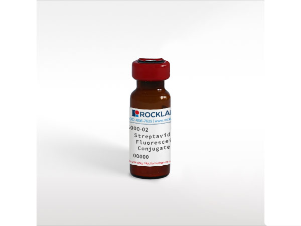
Background

Streptavidin is isolated from bacteria, Streptomyces avidinii, and has an exceptionally high binding affinity for B7 (biotin). Rockland offers streptavidin in unconjugated and conjugated forms for common immunoassays including ELISA, western blotting, immunohistochemistry. Streptavidin is a tetrameric protein capable of binding 4 biotin groups to each molecule of streptavidin. While streptavidin has identical binding properties as avidin, it lacks the glycoprotein portion of the molecule and therefore shows less non-specific binding. Streptavidin is a slightly smaller molecule with a molecular weight of approximately 53.6 kDa. The sequence of avidin only shows 30% homology with streptavidin, and anti-avidin and anti-streptavidin antibodies are not immunologically cross reactive. Rockland conjugates a broad group of secondary antibodies to many of the classic fluorescent markers including fluorescein, rhodamine, Texas Red, CyDyes™ and Phycoerythrin (RPE). All of the conjugates are ideal for various immunofluorescence based assays including fluorescent western blotting, immunofluorescence microscopy, FLISA, and more. Rockland also produces many next generation fluorochrome dyes designed for detection of primary antibodies in multiplex, multi-color analysis.

Product Details

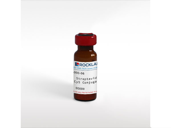
Streptavidin Cy5 Conjugated - S000-06
SA, S avidin, streptococcus avidin, Streptavidin Cy5 Conjugated, Cy5 conjugated Streptavidin
Cy5™

Target Details

Cy5 conjugated Streptavidin was prepared from chromatographically purified streptavidin followed by conjugation to the dye Cy5 and extensive dialysis against the buffer stated above.   Assay by immunoelectrophoresis resulted in a single precipitin arc against anti-Streptavidin.  No reaction was observed against anti-Avidin.

Application Details

Dot Blot, WB
ELISA, Functional Assay, IF, IHC, Multiplex  - View References
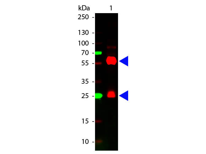
Streptavidin Cy5 Conjugated has been tested by dot blot and western blot and is designed for immunofluorescence microscopy, fluorescence based plate assays (FLISA) and fluorescent western blotting. This product is also suitable for multiplex analysis, including multicolor imaging, utilizing various commercial platforms.

Formulation

Lyophilized
1.0 mg/mL by UV absorbance at 280 nm
0.02 M Potassium Phosphate, 0.15 M Sodium Chloride, pH 7.2
0.01% (w/v) Sodium Azide
10 mg/mL Bovine Serum Albumin (BSA) - Immunoglobulin and Protease free
1.0 mL
Restore with deionized water (or equivalent)

Shipping & Handling

Ambient
Store vial at 4° C prior to restoration.   For extended storage aliquot contents and freeze at -20° C or below.  Avoid cycles of freezing and thawing.  Centrifuge product if not completely clear after standing at room temperature.  This product is stable for several weeks at 4° C as an undiluted liquid.  Dilute only prior to immediate use. 
Expiration date is one (1) year from date of receipt.
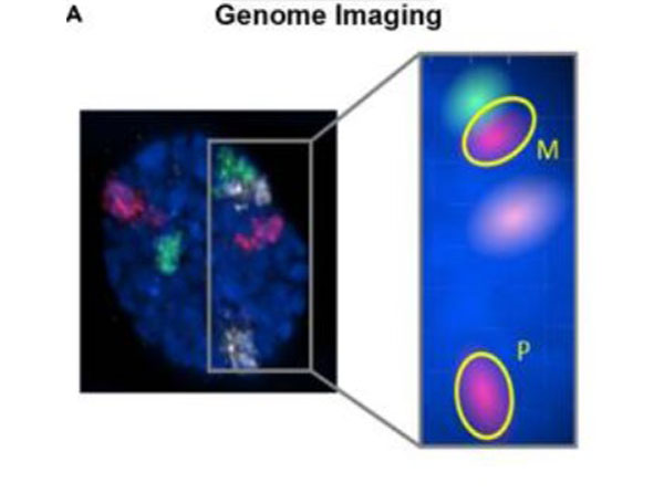
(). Functional organization of the maternal and paternal human 4D Nucleome. IScience
Applications
ISH, FISH
(). Voltage-gated calcium channel subunit α2δ-3 shapes light responses of mouse retinal ganglion cells mainly in low and moderate light levels. bioRxiv
Applications
Other
(). Snap chip for cross-reactivity-free and spotter-free multiplexed sandwich immunoassays. J Vis Exp.
Applications
Other
(). Novel glypican-3-binding peptide for in vivo hepatocellular carcinoma fluorescent imaging. Bioconjug Chem.
Applications
IHC, ICC, Histology
(). Retinal output changes qualitatively with every change in ambient illuminance. Nat Neurosci.
Applications
IF, Confocal Microscopy
(). Effects of the jimpy mutation on mouse retinal structure and function. J Comp Neurol.
Applications
IHC, ICC, Histology; Immuno-EM
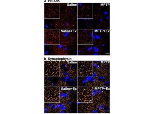
(). Treadmill exercise reverses dendritic spine loss in direct and indirect striatal medium spiny neurons in the 1-methyl-4-phenyl-1,2,3,6-tetrahydropyridine (MPTP) mouse model of Parkinson's disease. Neurobiology of Disease
Applications
IHC, ICC, Histology
(). Remodeling of three-dimensional organization of the nucleus during terminal keratinocyte differentiation in the epidermis. The Journal of Investigative Dermatology
Applications
ISH, FISH; IF, Confocal Microscopy
(). Antibody colocalization microarray: a scalable technology for multiplex protein analysis in complex samples. Molecular & Cellular Proteomics : Mcp
Applications
Microarray, Microarray chip
(). p63 regulates Satb1 to control tissue-specific chromatin remodeling during development of the epidermis. The Journal of Cell Biology
Applications
IF, Confocal Microscopy; Multiplex Assay

Invalid lot number

If you need help finding your CoA, contact us

This product is for research use only and is not intended for therapeutic or diagnostic applications. Please contact a technical service representative for more information. All products of animal origin manufactured by Rockland Immunochemicals are derived from starting materials of North American origin. Collection was performed in United States Department of Agriculture (USDA) inspected facilities and all materials have been inspected and certified to be free of disease and suitable for exportation. All properties listed are typical characteristics and are not specifications. All suggestions and data are offered in good faith but without guarantee as conditions and methods of use of our products are beyond our control. All claims must be made within 30 days following the date of delivery. The prospective user must determine the suitability of our materials before adopting them on a commercial scale. Suggested uses of our products are not recommendations to use our products in violation of any patent or as a license under any patent of Rockland Immunochemicals, Inc. If you require a commercial license to use this material and do not have one, then return this material, unopened to: Rockland Inc., P.O. BOX 5199, Limerick, Pennsylvania, USA.