Enhanced HCP Coverage Analysis utilizing Multiplexed 2D Electrophoresis
A crucial prerequisite in the drug manufacturing process is an efficient analysis of Host Cell Protein (HCP) impurities that result from process specific expression conditions as well as downstream purification procedures. Present guidelines call for minimum levels of HCP contaminants left behind during the purification process from the expression hosts. To investigate the presence of residual contamination of the final biopharmaceutical product, the development of polyclonal antibodies with maximum coverage against native HCP lysate provides a valuable tool to demonstrate product purity.
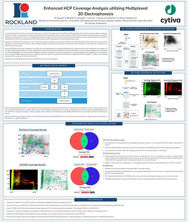
Quality of the HCP specific antibody is most commonly analyzed via ELISA and orthogonal methods like 1D and 2D-PAGE assays where the coverage of the antibody is confirmed. Determining anti-HCP antibody coverage is essential to ascertaining the robustness of the antibody in comprehensive reactivity toward HCPs during process points.
Here we evaluate the coverage of a generic anti-CHO-HCP antibody (Rockland Inc.) by 2D-PAGE separation using two experimental methods for the analysis: (A) chemiluminescent based detection method; and (B) Two-dimensional Differential In Blot Electrophoresis (2D-DIBE). For (A): the proteins detected by the antibody on a Western Blot (WB) via a chemiluminescent detection system, are equated to the proteins separated on a separate SDS-gel and visualized by an in-gel protein stain. For (B): 2D-DIBE utilizes a fluorescent multiplexing approach where the proteins and antibodies are tagged with different CyDyes™. Proteins and antibody coverage can be analyzed on the same membrane.
Sensitivity and antibody reactivity of the two methods are compared and analyzed.