Datasheet is currently unavailable. Try again or CONTACT US
Datasheet
$449.00 /Per Item
Availability: Ships next business day
Shipping info:

$50.00 to US & $70.00 to Canada for most products. Final costs are calculated at checkout.

Background

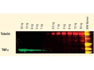
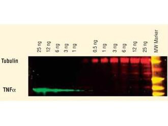
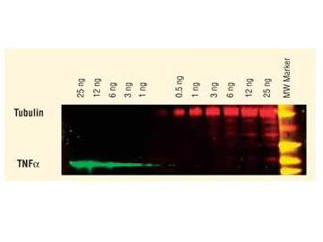
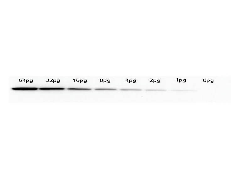
GFP Dylight™ 488 Conjugated Antibody is designed for immunofluorescence microscopy, fluorescence based plate assays (FLISA) and fluorescent western blotting. This product is also suitable for multiplex analysis, including multicolor imaging, utilizing various commercial platforms.

Product Details

Anti-GFP (GOAT) Antibody DyLight™ 488 Conjugated (Min X Hu Ms & Rt Serum Proteins) - 600-141-215
goat anti-GFP Antibody DyLight™ 488 Conjugation, DyLight™ 488 conjugated goat anti-GFP antibody, Green Fluorescent Protein, GFP antibody, Green Fluorescent Protein antibody, EGFP, enhanced Green Fluorescent Protein, Aequorea victoria, Jellyfish
Goat
DyLight™ 488
Polyclonal
IgG

Target Details

GFP, eGFP, rGFP
Recombinant Protein
The immunogen is a Green Fluorescent Protein (GFP) fusion protein corresponding to the full length amino acid sequence (246aa) derived from the jellyfish Aequorea victoria.
GFP Dylight™ 488 Conjugated Antibody was prepared from monospecific antiserum by immunoaffinity chromatography using Green Fluorescent Protein (Aequorea victoria) coupled to agarose beads followed by solid phase adsorption(s) to remove any unwanted reactivities.   Assay by immunoelectrophoresis resulted in a single precipitin arc against anti-Goat Serum and purified and partially purified Green Fluorescent Protein (Aequorea victoria).  No reaction was observed against Human, Mouse or Rat serum proteins.

Application Details

Dot Blot, WB
FC, IF, IHC, IP, Multiplex  - View References
Anti-GFP Dylight™ 488 Conjugated Antibody has been tested by dot blot and western blot. The emission spectra for this DyLight™ conjugate match the principle output wavelengths of most common fluorescence instrumentation.

Formulation

Lyophilized
1.0 mg/mL by UV absorbance at 280 nm
0.02 M Potassium Phosphate, 0.15 M Sodium Chloride, pH 7.2
0.01% (w/v) Sodium Azide
10 mg/mL Bovine Serum Albumin (BSA) - Immunoglobulin and Protease free
100 µL
Restore with deionized water (or equivalent)

Shipping & Handling

Ambient
Store Conjugated anti-GFP at 4° C prior to restoration.   For extended storage aliquot contents and freeze at -20° C or below.  Avoid cycles of freezing and thawing.  Centrifuge product if not completely clear after standing at room temperature.  This product is stable for several weeks at 4° C as an undiluted liquid.  Dilute only prior to immediate use.
Expiration date is one (1) year from date of receipt.
(). The Effect of Aging on Retinal Function and Retinal Ganglion Cell Morphology Following Intraocular Pressure Elevation. Frontiers in Aging Neuroscience
Applications
IHC, ICC, Histology
(). Macrophage network dynamics depend on haptokinesis for optimal local surveillance. ELife
Applications
IF, Confocal Microscopy; TUNEL
(). Retinal ganglion cell dysfunction in mice following acute intraocular pressure is exacerbated by P2X7 receptor knockout. Sci Rep.
Applications
IHC, ICC, Histology
(). Generation of human pluripotent stem cell-derived fused organoids with oligodendroglia and myelin. STAR Protoc.
Applications
IF, Confocal Microscopy
(). Establishment of a fluorescent reporter of RNA-polymerase II activity to identify dormant cells. Nat Commun.
Applications
IF, Confocal Microscopy
(). Inhibition of HIV infection by structural proteins of the inner nuclear membrane is associated with reduced chromatin dynamics. Cell Rep.
Applications
IF, Confocal Microscopy; WB, IB, PCA
(). Macrophage network dynamics depend on haptokinesis for optimal local surveillance. bioRxiv Preprint
Applications
IF, Confocal Microscopy
(). The N-Terminal Domain of cGAS Determines Preferential Association with Centromeric DNA and Innate Immune Activation in the Nucleus. Cell Reports
Applications
IF, Confocal Microscopy
(). A subset of skin macrophages contributes to the surveillance and regeneration of local nerves. Immunity
Applications
IF, Confocal Microscopy
(). OLIG2 drives abnormal neurodevelopmental phenotypes in human iPSC-based organoid and chimeric mouse models of down syndrome. Cell Stem Cell.
Applications
FC, FACS, FLOW; IF, Confocal Microscopy
(). Fractionation enhances acute oligodendrocyte progenitor cell radiation sensitivity and leads to long term depletion. Glia
Applications
IHC, ICC, Histology
(). Viral targeting of TFIIB impairs de novo polymerase II recruitment and affects antiviral immunity. PLOS Pathogens
Applications
IP, Co-IP; WB, IB, PCA
(). Ganglion Cell Assessment in Rodents with Retinal Degeneration. Methods Mol Biol.
Applications
IF, Confocal Microscopy
(). Distinct myeloid cell subsets promote meningeal remodeling and vascular repair after mild traumatic brain injury. Nat Immunol.
Applications
IHC, ICC, Histology
(). Allele-Specific Silencing of Mutant mRNA Rescues Ultrastructural and Arrhythmic Phenotype in Mice Carriers of the R4496C Mutation in the Ryanodine Receptor Gene ( RYR2). Circ Res.
Applications
IF, Confocal Microscopy
(). Direct Correlation between Motile Behavior and Protein Abundance in Single Cells. PLOS Computational Biology
Applications
WB, IB, PCA
(). Direct correlation between motile behavior and protein abundance in single cells. PLoS Comput Biol.
Applications
WB, IB, PCA
(). New insights into lineage restriction of mammary gland epithelium using parity-identified mammary epithelial cells. Breast Cancer Res.
Applications
IF, Confocal Microscopy; Multiplex Assay
(). Immunolocalization of the P2X4 receptor on neurons and glia in the mammalian retina. Neuroscience.
Applications
IF, Confocal Microscopy; Multiplex Assay
(). OLIG gene targeting in human pluripotent stem cells for motor neuron and oligodendrocyte differentiation. Nat Protoc.
Applications
IF, Confocal Microscopy; Multiplex Assay

Invalid lot number

If you need help finding your CoA, contact us

This product is for research use only and is not intended for therapeutic or diagnostic applications. Please contact a technical service representative for more information. All products of animal origin manufactured by Rockland Immunochemicals are derived from starting materials of North American origin. Collection was performed in United States Department of Agriculture (USDA) inspected facilities and all materials have been inspected and certified to be free of disease and suitable for exportation. All properties listed are typical characteristics and are not specifications. All suggestions and data are offered in good faith but without guarantee as conditions and methods of use of our products are beyond our control. All claims must be made within 30 days following the date of delivery. The prospective user must determine the suitability of our materials before adopting them on a commercial scale. Suggested uses of our products are not recommendations to use our products in violation of any patent or as a license under any patent of Rockland Immunochemicals, Inc. If you require a commercial license to use this material and do not have one, then return this material, unopened to: Rockland Inc., P.O. BOX 5199, Limerick, Pennsylvania, USA.