Background

Anti-Rabbit IgG (H&L) DyLight 488 Antibody generated in donkey detects reactivity to Rabbit IgG. Secreted as part of the adaptive immune response by plasma B cells, immunoglobulin G constitutes 75% of serum immunoglobulins. Immunoglobulin G binds to viruses, bacteria, as well as fungi and facilitates their destruction or neutralization via agglutination (and thereby immobilizing them), activation of the compliment cascade, and opsonization for phagocytosis. The whole IgG molecule possesses both the F(c) region, recognized by high-affinity Fc receptor proteins, as well as the F(ab) region possessing the epitope-recognition site. Both the Heavy and Light chains of the antibody molecule are present. Secondary Antibodies are available in a variety of formats and conjugate types. When choosing a secondary antibody product, consideration must be given to species and immunoglobulin specificity, conjugate type, fragment and chain specificity, level of cross-reactivity, and host-species source and fragment composition.

Product Details

Donkey Anti-
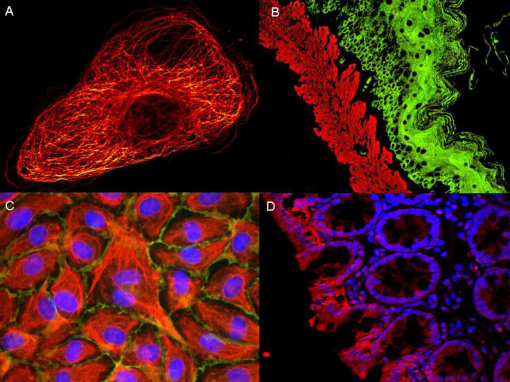
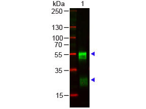
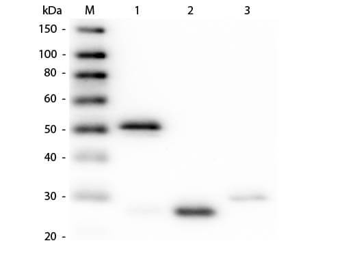
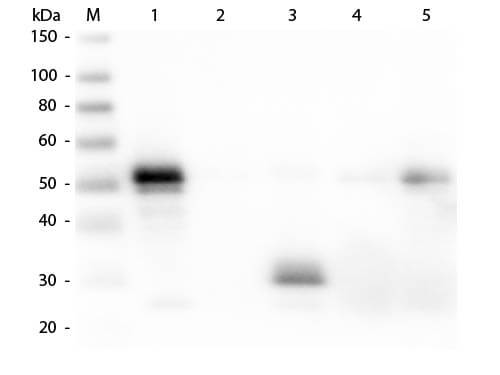
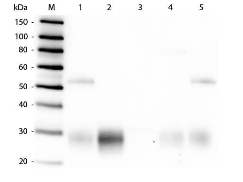
Rabbit IgG (H&L) Antibody DyLight™ 488 Conjugated (Min X Bv Ch Gt GP Ham Hs Hu Ms Rt & Sh Serum Proteins) - 611-741-127
Donkey Anti-Rabbit IgG Antibody DyLight 488™ Conjugated, Donkey Anti Rabbit IgG DyLight 488™ Conjugated Antibody
Donkey
IgG (H&L)
DyLight™ 488
Polyclonal
IgG

Target Details

Rabbit
Rabbit IgG whole molecule
This product was prepared from monospecific antiserum by immunoaffinity chromatography using Rabbit IgG coupled to agarose beads followed by solid phase adsorption(s) to remove any unwanted reactivities.  Assay by immunoelectrophoresis resulted in a single precipitin arc against anti-Donkey Serum, Rabbit IgG and Rabbit Serum.  No reaction was observed against Bovine, Chicken, Goat, Guinea Pig, Hamster, Horse, Human, Mouse, Rat and Sheep Serum Proteins.  This antibody will react with heavy chains of rabbit IgG and with light chains of most rabbit immunoglobulins.

Application Details

Dot Blot, WB
IF, IHC, Multiplex  - View References
Anti-Rabbit IgG (H&L) DyLight 488 Antibody has been tested by dot blot and western blot and is designed for immunofluorescence microscopy, fluorescence based plate assays (FLISA) and fluorescent western blotting. This product is also suitable for multiplex analysis, including multicolor imaging, utilizing various commercial platforms. The emission spectra for this DyLight™ conjugate match the principle output wavelengths of most common fluorescence instrumentation.

Formulation

Lyophilized
1.0 mg/mL by UV absorbance at 280 nm
0.02 M Potassium Phosphate, 0.15 M Sodium Chloride, pH 7.2
0.01% (w/v) Sodium Azide
10 mg/mL Bovine Serum Albumin (BSA) - Immunoglobulin and Protease free
100 µL
Restore with deionized water (or equivalent)

Shipping & Handling

Ambient
Store vial at 4° C prior to restoration.   For extended storage aliquot contents and freeze at -20° C or below.  Avoid cycles of freezing and thawing.  Centrifuge product if not completely clear after standing at room temperature.  This product is stable for several weeks at 4° C as an undiluted liquid.  Dilute only prior to immediate use. 
Expiration date is one (1) year from date of receipt.
(). Dietary carbohydrate influences the colocalization pattern of Glucagon-like Peptide-1 with neurotensin in the chicken ileum. Domest Anim Endocrinol.
Applications
Undefined
(). Dietary carbohydrate modifies the density of L cells in the chicken ileum. J Vet Med Sci.
Applications
IF, Confocal Microscopy
(). Citrullination regulates wound responses and tissue regeneration in zebrafish. J Cell Biol.
Applications
IF, Confocal Microscopy
(). Novel IQCE variations confirm its role in postaxial polydactyly and cause ciliary defect phenotype in zebrafish. Hum Mutat.
Applications
IF, Confocal Microscopy; IHC, ICC, Histology
(). Glucagon-like peptide-1 is co-localized with neurotensin in the chicken ileum. Cell Tissue Res.
Applications
IF, Confocal Microscopy; Multiplex Assay
(). In vivo micro-scale tomography of ciliary behavior in the mammalian oviduct. Scientific Reports
Applications
IF, Confocal Microscopy; Multiplex Assay
(). Distinct distribution of the tensin family in the mouse kidney and small intestine. Exp Anim.
Applications
IF, Confocal Microscopy
(). ETS1 regulates the expression of ATXN2. Hum Mol Genet.
Applications
IF, Confocal Microscopy; Multiplex Assay

Invalid lot number

If you need help finding your CoA, contact us

This product is for research use only and is not intended for therapeutic or diagnostic applications. Please contact a technical service representative for more information. All products of animal origin manufactured by Rockland Immunochemicals are derived from starting materials of North American origin. Collection was performed in United States Department of Agriculture (USDA) inspected facilities and all materials have been inspected and certified to be free of disease and suitable for exportation. All properties listed are typical characteristics and are not specifications. All suggestions and data are offered in good faith but without guarantee as conditions and methods of use of our products are beyond our control. All claims must be made within 30 days following the date of delivery. The prospective user must determine the suitability of our materials before adopting them on a commercial scale. Suggested uses of our products are not recommendations to use our products in violation of any patent or as a license under any patent of Rockland Immunochemicals, Inc. If you require a commercial license to use this material and do not have one, then return this material, unopened to: Rockland Inc., P.O. BOX 5199, Limerick, Pennsylvania, USA.