Tips for Flow Cytometry
-
Reducing noise from your fluorescent signal
A critical step during optimization of flow cytometry experiments is reagent (antibody) titration. For optimal results, you must determine the minimum amount of antibody required to achieve antigen-binding saturation. This will help you improve the specificity and intensity of your fluorescent signal while minimizing background. If you are simultaneously staining with multiple colors, this titration step will also allow you to detect unexpected cross-reactivity between primary and labeled secondary antibodies.
-
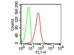
Choosing the right controls for flow cytometry
Always remember to include negative controls of the same isotype as the labeled antibody so that you can determine the extent of background signal in your experiments. This is true when staining with a labeled primary antibody only or when using a combination of primary antibody and phycoerythrin-labeled secondary antibody. For best results remember to always include a sample of unstained cells (incubated in parallel with your stained samples) so that you can control for background derived from auto-fluorescence. Whenever possible, include cells known to express the antigen of interest, as well as cells known to lack the same protein since they will help to determine the specificity of the antibodies used.
-
Optimize permeabilization and fixation to improve detection of intracellular proteins
Intracellular targets entail a more challenging detection since they have additional requirements beyond antibody specificity, including the ability to successfully cross the permeable cell membrane. Optimization of cell fixation and permeabilization is a critical parameter that needs to be empirically determined in order to balance the integrity of the intracellular structures with cell permeability. Always use freshly prepared solutions of high-purity paraformaldehyde for fixation and initially try mild detergents (i.e., Tween 20) for permeabilization. If your fixation and permeabilization steps need further improvement, try increasing the formaldehyde concentration gradually up to 4% or try ethanol or methanol and using other detergents including Triton X-100, or saponin without fixation or alcohol fixation.
-
Stain dead cells to obtain meaningful data from viable cells
Since dead cells can bind non-specifically to any antibody, it is imperative to keep those out of the analysis. The best way to do this is by using a fluorescent dye that will pass through damaged plasma membranes and thus stain dead cells. In most cases and especially when performing staining of intracellular targets, it is advisable to use dyes that covalently bind and stain non-viable cells so that they won't leak out once the cell is permeabilized.
-
Keeping fluorescent signal at high intensity
When performing staining of cell surface markers always consider the possibility that extracellular antigens can be internalized upon antibody binding. This naturally occurring phenomenon can have a significant negative effect on the intensity of your fluorescent signal and can be prevented by:
- Working with aggregate-free antibody solutions
- Keeping your samples ice-cold or refrigerated
- Including sodium azide in the staining buffer so that cellular metabolic activity is down-regulated during staining. Also, consider using monovalent antibodies in the form of F(ab) fragments.