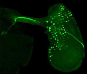
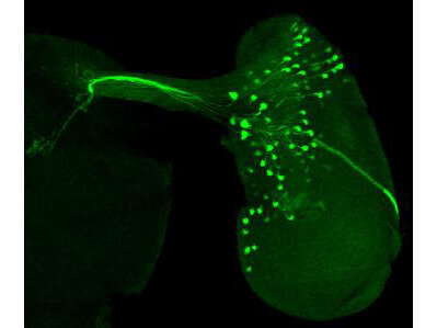
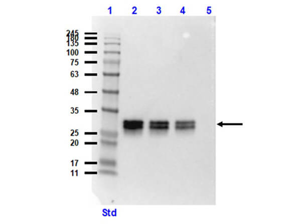
Green fluorescent protein (GFP) has revolutionized the field of molecular and cell biology as a valuable tool for tracking protein expression and localization in living cells. GFP is a 27 KDa cytoplasmic protein that was first purified from the jellyfish Aequorea victoria. Consistent with other fluorescent proteins, the fluorophore of GFP is formed by autocatalysis from three consecutive amino acid residues within a polypeptide chain. To maximize the utility of GFP in certain applications such as western blotting, immunoprecipitation, and fluorescent microscopy, researchers require reliable and specific GFP antibodies.
This article will provide a detailed overview of GFP antibodies from chicken, goat, mouse, and rabbit hosts, and how they can address different concerns in scientific research.
GFP Variants
Various GFP variants have been developed to improve upon the limitations of wild-type GFP, such as lower brightness, fluorescence intensity, and usability in specific experiments. For example, the mutation of Ser65 to Thr (S65T) simplifies the excitation spectrum to a single peak at around 490 nm of enhanced amplitude.1 Enhanced GFP (eGFP) has a fluorescence 6 times brighter than that of wild-type GFP, faster maturation, and is less sensitive to temperature-dependent misfolding. On the other hand, eGFP and S65T-GFP photobleach about twice as fast as wild-type GFP.2 Another variant, RS-GFP, has been engineered to have a slightly red-shifted spectrum, making it a valuable tool for FRET experiments. This also applies to yellow fluorescent protein (YFP), which emits yellow light instead of green.
To summarize, GFP variants allow fine-tuning protein expression and localization studies. Regarding the specificity of GFP antibodies, the differences are negligible since the variants differ only in a few amino acids.
Comparison of GFP Variants
| Variant | Excitation (nm) | Emission (nm) | Extinct. Coeff. (M-1cm-1) | Quantum yield (Φ) | Brightness (nM-1cm-1) | Maturation (min) |
| GFP, rGFP | 395 (475) | 509 | 25,000 | 0.79 | 19.75 | 36 min |
| eGFP | 488 | 507 | 55,900 | 0.60 | 33.54 | 25 min |
| S65T-GFP | 490 | 510 | 55,000 | 0.64 | 35.2 | - |
| RS-GFP | 493 | 510 | 47,000 | 0.36 | 16.92 | 180 min |
| YFP | 513 | 527 | 67,000 | 0.67 | 44.89 | 9 min |
Data retrieved from FPbase3
Monoclonal vs. Polyclonal GFP Antibodies
Monoclonal GFP antibodies are generated from a single clone of B cells, producing identical antibodies that specifically bind to a single epitope. In contrast, polyclonal GFP antibodies are purified from serum, producing a heterogeneous mixture of antibodies that recognize multiple epitopes.
The main advantage of monoclonals is their high specificity and consistency, as lot-to-lot variation is minimal. However, the use of polyclonal antibodies can be advantageous because the heterogeneous binding of several different epitopes makes it more likely that these reagents will successfully bind GFP in a variety of different assay conditions and immunoassays. While in some applications (i.e. Western blot) denatured protein must be detected, other applications require the detection of native GFP. Polyclonal antibodies are usually more capable of detecting both variants.
Since all anti-GFP antibodies presented here are validated and tested in numerous applications, one should make the purchase decision based on the specific product references and applications.
GFP Antibodies from Different Host Species
GFP antibodies from different host species, such as mouse, rabbit, goat, and chicken, offer distinct advantages in various applications. The majority of the dissimilarities arise from variations in structure and antigenicity.
With the exception of avian GFP antibodies, which are typically IgY, other common GFP antibodies are from the immunoglobulin G class. One key structural difference between IgY and IgG antibodies lies in their respective heavy chains. IgG antibodies have three constant regions in their heavy chains, whereas IgY antibodies have four, which increases the MW of IgY to 180 kDa (see figure). The absence of the hinge region in IgY leads to increased stiffness, which makes it more resistant to proteolytic degradation and generally more stable than IgG. Other structural variations and phylogenetic distance result in non-reactivity to specific components of the human immune system such as Fc receptors, making it an ideal tool when minimal interference is desired.4

Figure: Structure of IgG and IgY
In cases where a preference for a rabbit or goat antibody arises, such as when a secondary antibody is already being utilized, the selection should be guided by manufacturer-tested or literature-cited (suggested) applications. Antibodies from different host species can be advantageous in multiplex experiments. For example, combining the detection of an antigen with a mouse primary antibody and a GFP antibody from goat. This allows for the detection of two different targets in the same sample using different labels. Multiplex experiments can increase the efficiency of experiments and save time and resources by allowing for the simultaneous detection of multiple targets.
Features and Benefits of Goat, Rabbit, Chicken, and Mouse Antibodies
| Host Species | Goat | Rabbit | Chicken | Mouse |
| Immunoglobulin Class: | IgG | IgG | IgY | IgG |
| Format: | Polyclonal | Polyclonal, Monoclonal | Polyclonal | Monoclonal |
| Source: | Serum | Serum, cell culture supernatant | Serum, egg yolk | Cell culture supernatant |
| Molecular Weight: | 150 kDa | 150 kDa | 180 kDa | 150 kDa |
| Constant Domains (Heavy Chain): | 3 | 3 | 4 | 3 |
| Hinge Region: | Yes | Yes | No | Yes |
| pH Stability: | 2.0–11.0 | 2.0–11.0 | 3.5–11.0 | 2.0–11.0 |
| Heat Stability: | <75°C | <75°C | <70 °C | <75°C |
| Proteolytic Degradation: | Pepsin, papain, trypsin and chymotrypsin | Pepsin, papain, trypsin and chymotrypsin | Pepsin and papain | Pepsin, papain, trypsin and chymotrypsin |
| Complement Binding: | Yes | Yes | No | Yes |
| Fc Receptor Binding: | Yes | Yes | No | Yes |
| Binding to Protein A/G: | Low | High | No | High |
Table adapted from Pereira et al., 2019
Conclusion
GFP antibodies from different host species possess unique features that make them ideal for certain applications. The choice of GFP antibody depends on various factors, including the preferred clonality, the host species, and the intended use of the antibody. Product references play a crucial role in this decision-making process, as they provide valuable information about the performance and reliability of specific GFP antibodies. By referring to product references, one can gain insights into the antibody's performance in relevant experiments, helping you make informed choices. By choosing the right GFP antibody for a specific application, you can enhance the accuracy and efficiency of your experiments.
Recommended GFP Antibodies
References
- Ormö, M., Cubitt, A. B., Kallio, K., Gross, L. A., Tsien, R. Y., & Remington, S. J. (1996). Crystal structure of the Aequorea victoria green fluorescent protein. Science (New York, N.Y.), 273(5280), 1392–1395.
- Patterson, G. H., Knobel, S. M., Sharif, W. D., Kain, S. R., & Piston, D. W. (1997). Use of the green fluorescent protein and its mutants in quantitative fluorescence microscopy. Biophysical journal, 73(5), 2782–2790.
- Lambert T. J. (2019). FPbase: a community-editable fluorescent protein database. Nature methods, 16(4), 277–278.
- Michael, A., Meenatchisundaram, S., Parameswari, G., Subbraj, T., Selvakumaran, R., & Ramalingam, S. (2010). Chicken egg yolk antibodies (IgY) as an alternative to mammalian antibodies. Indian J. Sci. Technol, 3(4), 468-474.
- Pereira, E. P. V., van Tilburg, M. F., Florean, E. O. P. T., & Guedes, M. I. F. (2019). Egg yolk antibodies (IgY) and their applications in human and veterinary health: A review. International immunopharmacology, 73, 293–303.